Human Eye Anatomy and Basic Eye Facts of Biology, Chemistry and Physics

Eye Anatomy Structure, All Basic Structure
Anatomy and Parts of the Eye
The Human Eye is an Organ
This organ of the body is made up of different tissues which are composed of cells. Cells are the smallest, complete, whole parts of any organism as shown above in a diagram of the eukaryotic cell. Thus, the human eye is an organ of various tissues and cells that together assist in vision.
Important Eye Parts Include
This parts of the eye work to make the whole eye a funtioning organ: conjunctiva, cornea, pupil, iris, aqueous humor, lens, ciliary body and muscles, vitreous humor, retina, choroid, sclera, and optic nerve. The simpler diagram shown here first will highlight the basic, simpler features.. The conjunctiva is not shown in the eye figure, but it is the tissue that lines the outside of the eyeball (just outside the sclera) and the inside of the eyelids. A good way to remember the conjunctiva is to recall the itchy and red eye disease called conjunctivitis that occurs when the conjunctiva becomes irritated or inflammed. Conjunctivitis is a burning, irritating condition that causes reddening of the lining.
Notice the clear cornea and the lens. The fluid between the cornea and lens that is watery-clear, and called the aqueous humor. The ciliary body is part of the choroid layer, and it is able to cause the iris, which is also in the choroid, to expand and contract when assisted by the ciliary muscles. The pupil is the name for opening of the iris that dilates or contracts. The iris colors range from brown, blue, green, or other shades, and the pupil appears almost black. Anyone can check their eyes in the mirror and see the light reflex induced by changes in light intensity. Shine a light in one pupil, and then away and back again. See the contraction and expansion of the pupil as light is first shone, and then taken away from the pupil. This response is automatic (part of the autonomic system).
Notice the optic nerve. The optic nerve carries electrical signals, generated by the light-induced chemical changes, to sight areas of the brain, that interpret what the signals mean, and this enables people to see. Both nerves and internal blood vessels are channeled through this area, and since there are no retinal receptors here it is a blind spot -- a small, but real area of the retina.
Both the cornea and the lens bend entering light rays, and focus them on the retina, which is in the back (posterior) region of the eye. The inner fluid of the eye, between the lens and the retina, is vitreous fluid. This vitreous fluid is jelly-like and clear and gives the eye shape and form, and keeps the retina pressed tightly against the choroid layer.
The sclera is the white covering of thick, protective tissue and the cornea is the clear extension of the sclera. When people see the eyes of others the white part of the eye is the sclera. Note that the cornea is a clear extension, or part of the white sclera. The cornea is clear and focuses light rays in conjunction with the lens. With age, the cornea may become cloudy and require surgical intervention. The fovea or macula area is the most photoreceptor-concentrated region, and it also the most sensitive and important area of the retina for image formation and clarity.
Chemistry and Physics of the Eye, Rhodopsin, Cones and Nerve Signals
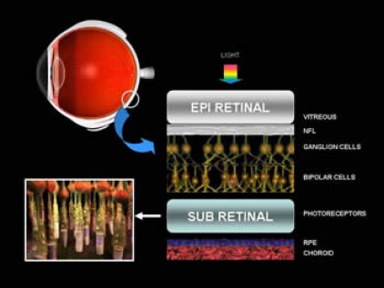
Retina, Ganglia (Nerve Cells), Bipolar Cells, Cones, Rods, Choroid Layer
The light rays are focused by the cornea and lens which cause light refraction onto the retina. The visible light radiations penetrate down into the photoreceptor regional which is subretinal (below the retina). The retina has ganglion cells and bipolar cells that interact with photoreceptor cones (color- and high- intensity, light-receptor cells; approximately 7 million cells) and rods (low intensity, light-receptor cells; approximately 120 million cells). These cells then transmit signals to the ganglia and bipolar cells and these signals are carried to the optic nerve and to the brain. Light reacts with rhodopsin found in both rods and cones. Notice the choroid layer lies beneath the retina and extends all the way around the eyeball. The chorion is enriched with blood vessles to nourish the eye, and the chorion is dark and pigmented, and prevents reflections inside the eye.
Eye, Vision Impairment Numbers and Satistics –14 Million People in the USA in 2006
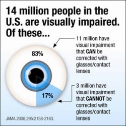
The Journal of the American Medical Association reported in 2006, that about 11 million have correctable vision problems, and some 3 million cannot be corrected. Yet, for those with vision problems, much can be done. There are simple glasses and contact lenses that can compensate for near-, far- sightedness and astigmatism. There is radial keratotomy with laser surgery to correct lens problems. And corneal transplants as well as lens implants make eye diseases correctable. Sometimes the retina becomes damaged, as in macular degeneration that affects the fovea of the retina. In retinosa pigmentosum there is progressive degenerative destruction of the retina with eventual complete loss of vision and blindness.
Eye Problem Corrections – Keratotomy, Glasses, Lenses, Implants

Myopia, Nearsightedness Corrected by Keratotomy
Imagine a movie screen that is blurry. The picture is out of focus. To get the image on the screeen the projectionist adjusts the lens of the projector. If the image falls in front of the screen, the lens is adjusted to make it fall back onto the screen. See the eye on the left with the focal point in front of the retina. The light rays are bent too much and they come to points way before the retina. If the lens is flattened out correctly, the image will fall onto the retina. Glasses and contact lenses can do that, and radial or laser keratotomy have the same effect. Keratatomy involves minor surgery that flattens the eye lens somewhat, and causes the image to form right on the retina. The vision problem is corrected without glasses or contact lenses.
VISIT Our Science Super School Store
All the Written Material within Site is Copyrighted 2010 and Owned by Dr. Donald Reinhardt, and this original material is protected legally by this copyright notice and by the Digital Millennium Act. None of this original material may be copied or reproduced without the expressed written consent of the author.
The author is a Freelance Science writer, and is available for specific assignments for those who are interested – by contacting adminstrator@sciencesuperchool.com. Other questions related to this teaching site should be directed to teacher@sciencesuperschool.com.